

Western blotting, also known as immunoblotting, is a powerful laboratory technique widely used in molecular biology, biochemistry, and immunology to detect specific proteins in a complex sample. By combining gel electrophoresis with antibody-based detection, western blotting allows researchers to not only confirm the presence of a protein but also analyze its size, abundance, and modifications. Understanding the procedure in detail is crucial for generating reliable and reproducible results.
1. Sample Preparation
The first step in the western blotting procedure involves preparing the protein samples. Cells or tissues are lysed using appropriate buffers containing detergents and protease inhibitors to prevent protein degradation. The total protein concentration is then quantified using assays such as the Bradford or BCA method. Proper sample preparation ensures that proteins remain intact and that their concentration is suitable for detection.
2. SDS-PAGE: Protein Separation
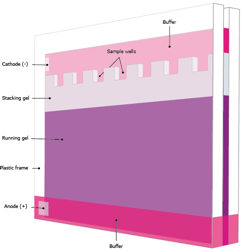
After preparation, protein transfer system are separated according to their molecular weight using sodium dodecyl sulfate-polyacrylamide gel electrophoresis (SDS-PAGE). SDS, a detergent, denatures proteins and imparts a negative charge proportional to their length, ensuring that proteins migrate solely based on size. The gel’s concentration is selected depending on the expected size of the target protein; higher concentrations resolve smaller proteins, while lower concentrations are better for larger proteins. Once loaded, an electric field drives the proteins through the gel, separating them into distinct bands.
3. Transfer to Membrane
Following electrophoresis, proteins are transferred from the gel onto a solid support, usually a nitrocellulose or polyvinylidene fluoride (PVDF) membrane. This step is critical because it makes the proteins accessible for antibody binding. Transfer can be performed using wet or semi-dry methods. In wet transfer, the gel and membrane are sandwiched between filter papers and immersed in transfer buffer while an electric current moves the proteins. Semi-dry transfer uses less buffer and is faster but may be less efficient for very large proteins. Proper transfer ensures that proteins are immobilized without distortion.
4. Blocking
To prevent nonspecific binding of antibodies, the membrane is incubated with a blocking solution. Common blockers include non-fat dry milk, bovine serum albumin (BSA), or commercial blocking agents. Blocking fills in the unoccupied sites on the membrane surface, reducing background noise and enhancing the specificity of the antibody-antigen interaction.
5. Primary Antibody Incubation
The membrane is then incubated with a primary antibody specific to the protein of interest. This antibody binds directly to the target protein. Incubation conditions, including antibody concentration, temperature, and duration, are optimized to maximize specific binding while minimizing nonspecific interactions. Overnight incubation at 4°C is common for sensitive detection.
6. Secondary Antibody Incubation
After washing away unbound primary antibodies, the membrane is exposed to a secondary antibody that recognizes the primary antibody. This secondary antibody is usually conjugated to an enzyme, such as horseradish peroxidase (HRP) or alkaline phosphatase (AP), which facilitates signal detection. The secondary antibody amplifies the signal, making it easier to detect low-abundance proteins.
7. Detection
Detection of the protein involves adding a substrate that reacts with the enzyme on the secondary antibody, producing a visible signal. Chemiluminescent substrates produce light that can be captured on X-ray film or with a digital imaging system, while colorimetric substrates generate a colored precipitate. The intensity of the signal correlates with the amount of protein present, allowing semi-quantitative analysis.
8. Data Analysis
Finally, the developed blot is analyzed to interpret the results. Bands corresponding to the protein of interest are compared to molecular weight markers to confirm their size. Densitometry software may be used to quantify protein levels, and loading controls such as β-actin or GAPDH are included to ensure equal sample loading.
Conclusion
Western blotting remains a cornerstone technique in biological research, providing a detailed and reliable method to study proteins. By carefully following each step—from sample preparation and SDS-PAGE to antibody detection and analysis—researchers can obtain precise insights into protein expression, modifications, and function. Mastery of western blotting procedure details ensures both reproducibility and accuracy in protein analysis.

Leave a Reply